Drawing strength from asymmetry!
Discovery of a new protein, PgfA, could be targeted for the development of new anti-TB drugs; writes Kuldeepkumar R Gupta

Tuberculosis (TB) kills more humans than any other disease caused by an infectious agent. Every year around 10 million people get infected with TB, and 1.4 million die because of it across the world. TB patients are treated with a cocktail of four antibiotics which target different processes in the TB-causing bacterium. Though an effective TB treatment is available, it takes at least six months for the treatment of drug-sensitive TB. Moreover, in recent years, there has been an increase in the cases of drug-resistant TB, which can take up to two years for the treatment. Such a prolonged treatment also causes several side effects such as weight loss, nausea, blurred vision, jaundice etc. Moreover, some TB patients may stop taking the medication because of side effects or lengthy treatment duration. This might cause the relapse of the disease and give rise to drug-resistant TB. Hence, there is an immediate need for effective TB treatments involving reduced pill burden and significantly shorter duration. TB is caused by the bacterium Mycobacterium tuberculosis, which belongs to a larger group of bacteria called mycobacteria. Interestingly, the leprosy-causing bacterium also belongs to this group. The bacteria responsible for Buruli ulcer and tuberculosis in birds and cattle also belong to mycobacteria group. Humans and chimpanzees are closely related species in terms of their DNA sequence. Likewise, the bacteria belonging to mycobacteria group have a very similar genetic material. Consequently, these closely related species have nearly identical physiological processes such as growth, division, and cell wall structure.
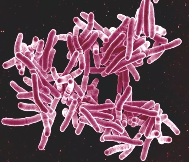
One way to develop novel therapeutics for TB treatment is to specifically target the indispensable proteins that are responsible for growth of Mycobacterium tuberculosis. Mycobacteria, like other bacteria, multiply by a process of binary fission, also known as cell division. In this process, the bacterial cell first grows for a certain time to increase its size by making new cell walls and cellular contents such as proteins, DNA, RNA etc. After the doubling of cellular contents, the cell divides in the middle to produce two daughter cells of equal size. Mycobacteria are rod-shaped bacteria, and they grow by adding new cell wall material at their poles. Hence, they exhibit the phenomenon of polar growth. Interestingly, one pole of the mycobacteria grows faster than the other pole. The fast-growing pole is called the old pole, while the slow growing pole is called the new pole. Because of this anomaly in the polar growth, the cell division produces two daughter cells of unequal cell lengths. The fast-growing pole or old pole produces a longer daughter cell, while slow-growing pole or new pole produces a shorter daughter cell. This phenomenon is referred to as asymmetric cell division, which produces variability in the size of cells in an otherwise genetically identical population. The heterogeneity in cell size imparts each cell differential susceptibility to antibiotics. Cells become sensitive to one kind of antibiotics and are resistant to others. Consequently, the variability caused by asymmetric cell division is partly responsible for the long duration of TB treatment. The asymmetric cell division is controlled by a protein called LamA. When the gene encoding LamA protein is deleted from the genome of mycobacteria, the cell division becomes relatively symmetric, and cells easily get killed by antibiotics. However, molecular mechanisms of asymmetric polar growth caused by LamA are still not known.
To answer this question, a team led by Indian origin scientist Kuldeepkumar R Gupta in the laboratory of Hesper Rego at Yale University School of Medicine used genetic, biochemical and microscopy approaches. The team discovered an unknown protein which interacts with LamA and assists it in carrying out asymmetric cell division. The team found that LamA guides this newly discovered protein called polar growth factor A (PgfA) to the fast-growing old pole. In the absence of PgfA, the old pole stops growing and cells eventually die. They also found that the cell death is caused by the destruction of the protective cell wall in the absence of PgfA. Through a series of experiments, Gupta and team subsequently showed that PgfA helps in the transport of a cell wall building block from inside of the cell to the site of cell wall synthesis at the poles.
Two out of four antibiotics used to treat TB target the cell wall of Mycobacterium tuberculosis. This cell wall structure is also conserved in other mycobacterial species. The mycobacterial cell wall is unique as it has multiple layers made up of sugars, peptides, and lipids. Apart from asymmetric cell division, this multi-layered cell wall is also partly responsible for the extreme antibiotic resistance shown by TB-causing bacteria. Thus, asymmetric cell division combined with unique cell wall structure confer mycobacteria a distinctive ability to withstand a very harsh antibiotic treatment. Discovery of PgfA connects the two important processes of cell division and cell wall synthesis in clinically important mycobacterial species. By targeting PgfA, one can target Achilles' heel of mycobacteria. Hence, PgfA represents a very viable target for the development of anti-TB drugs. Moreover, these drugs can also be used to treat other mycobacterial diseases since processes governed by PgfA are conserved across mycobacteria. The study has been published in the journal 'eLife', which is an open-access journal for members of the public. The title of the study is 'An essential periplasmic protein coordinates lipid trafficking and is required for asymmetric polar growth in mycobacteria'.
The writer is currently an associate research scientist in the Department of Microbial Pathogenesis at Yale University School of Medicine, New Haven, USA. Views expressed are personal


